Venous Thromboembolism – Diagnosis and Management
Effective Date: February 29, 2024
Recommendations and Topics
- Scope
- Key Recommendations
- Background And Epidemiology
- Risk Factors
- Assesment and Diagnosis
- Diagnosis of DVT
- Management of Acute DVT/PE
- Disgnosis of Pulmonary Embolis
- Supportive Measures
- Follow up
- Resources
Scope
This guideline provides recommendations for the diagnosis and management of venous thromboembolism (VTE) in adults aged ≥ 19 years with hemodynamic stability. It includes lower limb deep vein thrombosis (DVT) and pulmonary embolism (PE) diagnosis in the outpatient setting and management of acute VTE. Superficial thrombophlebitis and thrombosis in unusual sites (e.g., cerebral venous thrombosis, splanchnic vein thrombosis, upper extremity thrombosis) are outside the scope of this guideline. For information refer to the Thrombosis Canada Guidelines.
Key Recommendations1,2
- When DVT/PE is suspected, first calculate the Wells Score to determine the likelihood of DVT/PE as “likely” or “unlikely” before ordering any testing.
- For outpatients with suspected DVT/PE:
- Do not order D-dimer if DVT/PE is deemed “likely” per Wells Score. Proceed directly to imaging.
- Order D-dimer when deemed ‘unlikely’ per Wells Score because a negative test indicates imaging is not necessary and DVT/PE is excluded.
- For inpatients, proceed directly to imaging because risk stratification using D-dimer has not been validated.
- While awaiting objective imaging to diagnose VTE, start empiric anticoagulant therapy in patients with higher likelihood (“likely”) of DVT/PE.
- Most patients with hemodynamically stable VTE can be treated on an outpatient basis.
- Direct Oral Anticoagulants (DOACs) are considered as first line therapies for most outpatients. They are contraindicated in pregnancy, breastfeeding, liver failure (Child-Pugh class C), dialysis, or triple-positive antiphospholipid syndrome (i.e., has lupus anticoagulant, anticardiolipin and antibeta-2- glycoprotein-1 antibodies).
- Ensure appropriate anticoagulant dosage is used for the specific treatment phase (initial therapy, primary treatment, secondary prevention).
- Minimum duration of anticoagulation is 3-6 months for all patients with an acute DVT/PE.
- Referral to a thrombosis specialist is recommended to help determine optimal duration of anticoagulation. Continue anticoagulation therapy while awaiting referral.
- Avoid elective surgeries during the first 3-6 months of treatment.
- Hereditary thrombophilia testing and occult cancer screening are not indicated in most patients with thrombosis because results rarely influence management.
Background and Epidemiology
VTE affects at least one in 1000 individuals each year3 and is the third most common cause of vascular death worldwide.4 VTE most commonly manifests as DVT in the legs and/or PE.
Up to 10% of symptomatic PEs are fatal within the first hour of symptom onset.5 Independent predictors of mortality within the first few days after PE diagnosis include hypotension (systolic blood pressure < 90 mmHg), clinical right heart failure, right ventricular dilatation on computer tomography pulmonary angiogram (CTPA) or echocardiography, elevated troponin, and elevated brain natriuretic peptide.
Early diagnosis and treatment reduce VTE morbidity and mortality.
Risk Factors
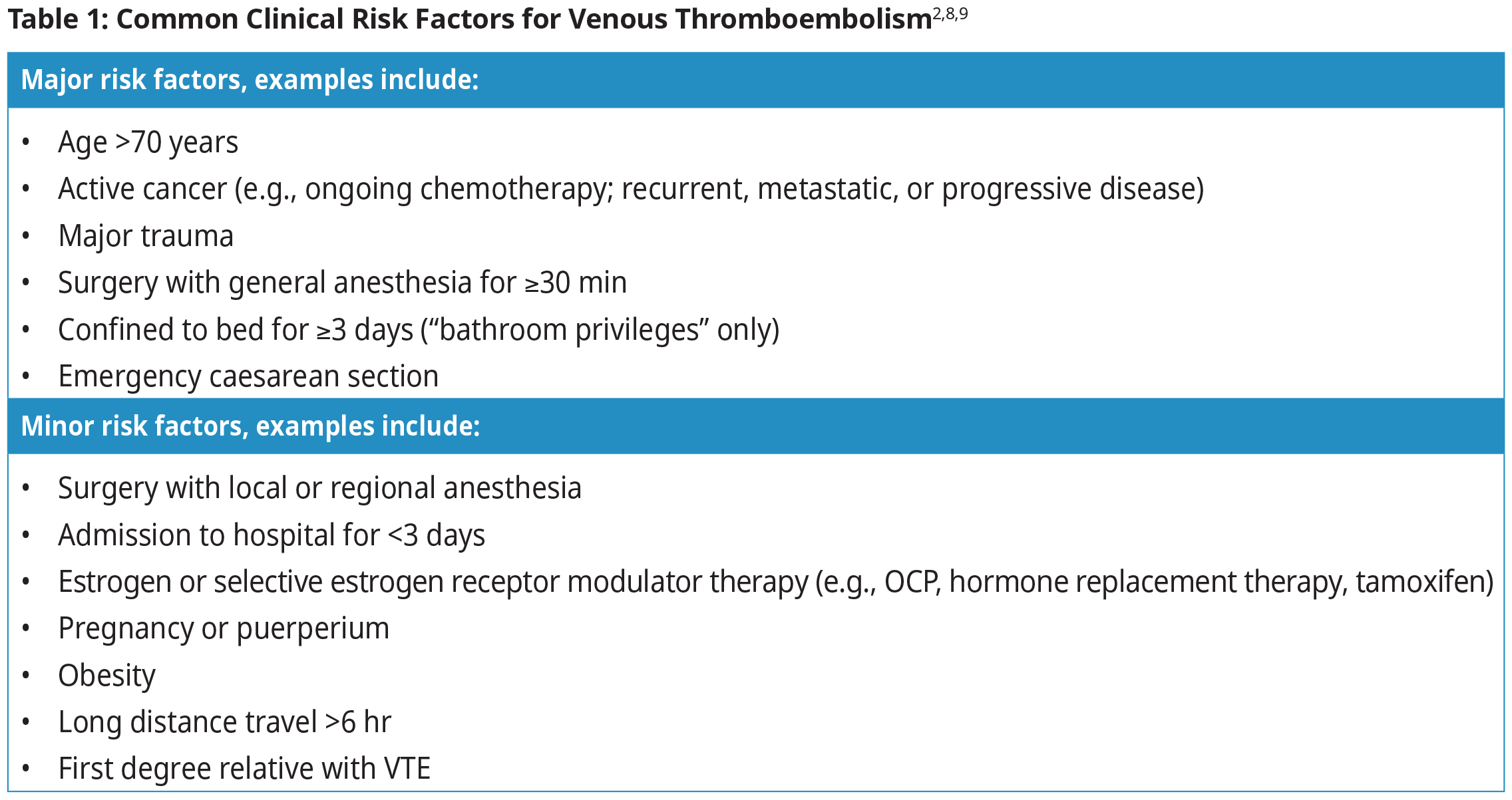
The most common VTE risk factors include recent surgery, hospitalization, active cancer, and high estrogen states such as oral contraception (OCP) and pregnancy.3,6,7 See Table 1: Common Clinical Risk Factors for Venous Thromboembolism for examples of major and minor VTE risk factors.
Assessment and Diagnosis
- Objective imaging and/or blood testing are required to confirm or exclude DVT/PE because signs and symptoms are nonspecific.
- Clinical pretest probability models (e.g., Wells scores) are recommended to help guide necessary investigations.
- Combined use of clinical pretest probability and D-dimer testing can help exclude VTE, but imaging tests are required to confirm a diagnosis.
- Patients with PE might have silent DVT, and some patients with DVT might have silent PE.
- Clinicians should always apply their clinical judgement and discretion in following the diagnostic algorithms, especially in cases where history or symptoms are not reliable and there are no alternative diagnoses to explain patient’s presentation.
Diagnosis of DVT
Classic signs and symptoms of DVT include limb swelling, pain or muscle ache, warmth, and erythema. Cyanotic discolouration or skin mottling might indicate extensive venous congestion from limb-threatening thrombosis. However, these findings are nonspecific for DVT.
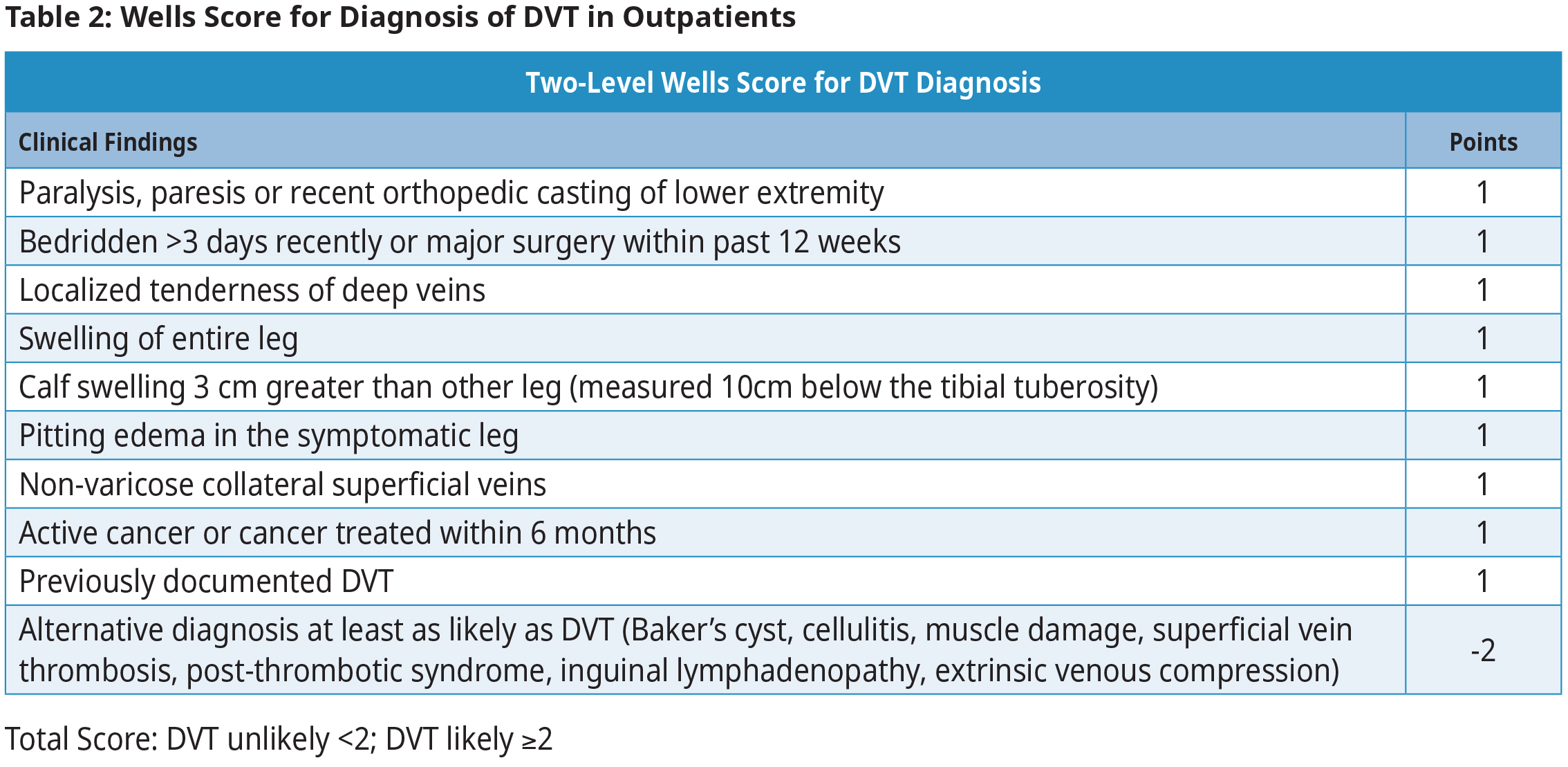
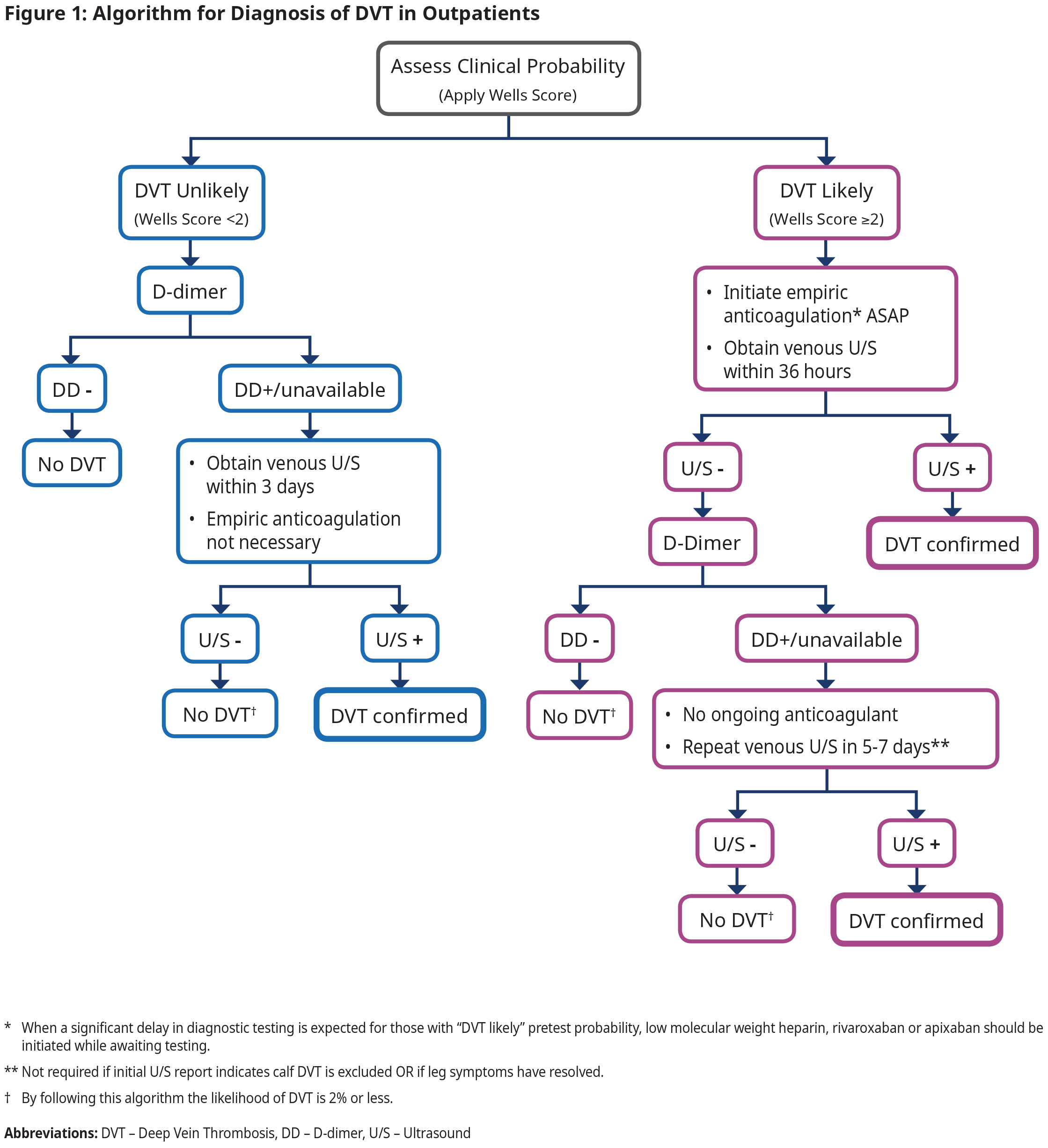
When a DVT is suspected, first calculate the Wells Score for DVT. See Table 2: Wells Score for Diagnosis of DVT in Outpatients. Total score indicates the probability of DVT as “unlikely” or “likely”. Follow the algorithm in Figure 1: Algorithm for Diagnosis of DVT in Outpatients to determine further investigations. For inpatients, proceed directly to imaging because risk stratification using D-dimer has not been validated.
While awaiting objective imaging to diagnose DVT, start empiric anticoagulant therapy in patients with higher likelihood (“likely”) of DVT.
Investigations used for diagnosis of DVT:
- Venous ultrasound – Various techniques, including compression and/or Doppler, are used. Order venous ultrasound of the affected limb(s) and specify that it is for possible DVT to ensure correct technique is used. See Appendix A: Proximal and Distal DVT for location of thrombus.
- D-dimer – This biomarker is elevated when there is active clot formation and breakdown. It can be elevated in acute DVT/PE, but also with infection, infarction, inflammation, surgery/trauma, cancer, and pregnancy. Consequently, an elevated result does not confirm DVT/PE, but a negative result can safely rule out an acute DVT in patients with “unlikely DVT” (See Figure 1: Algorithm for Diagnosis of DVT). See Appendix B: D-dimer Cutoff Values.
Diagnosis of Pulmonary Embolis
Classic signs and symptoms of PE include unexplained dyspnea, tachypnea, and tachycardia (common), pleuritic chest pain and hemoptysis (uncommon). Syncope, hypoxemia, hypotension, or other features of right ventricular dysfunction (e.g., distended jugular veins) suggest life-threatening thrombotic burden. However, all these findings are non-specific for PE.
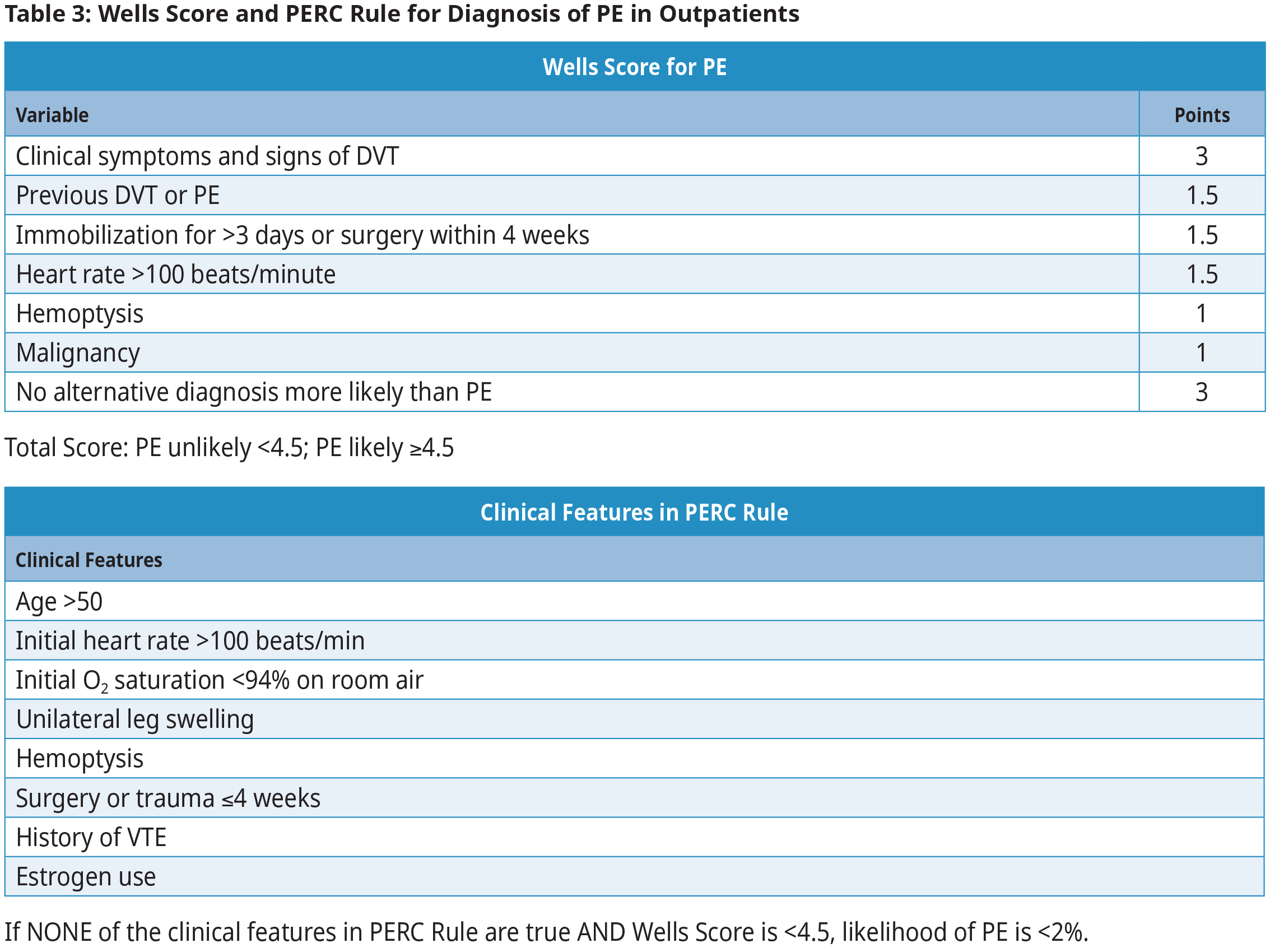
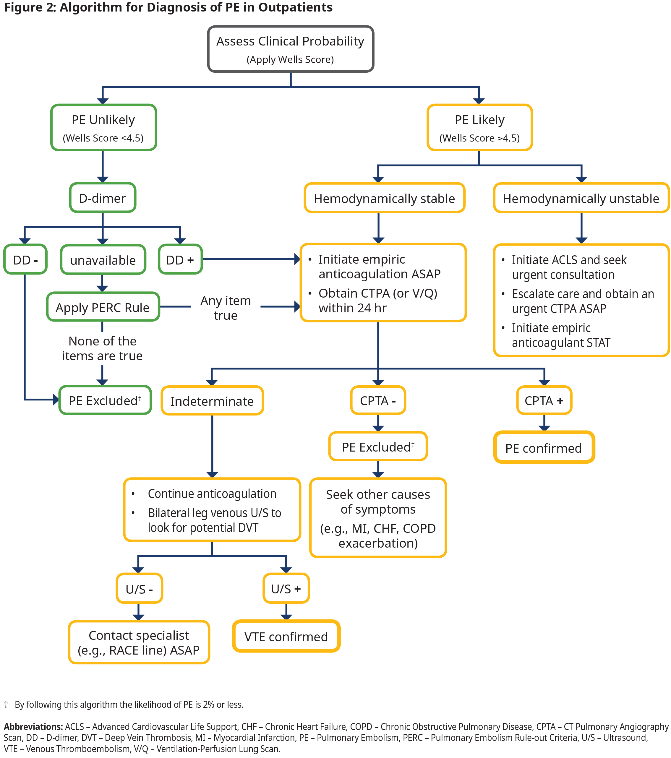
When a PE is suspected, first calculate the Wells Score for PE. See Table 3: Wells Score and PERC Rule for Diagnosis of PE in Outpatients. Total score indicates the probability of PE as “unlikely” or “likely”. Follow the algorithm in Figure 2: Algorithm for Diagnosis of PE in Outpatients to determine further investigations. For inpatients, proceed directly to imaging because risk stratification using D–dimer has not been validated.
While awaiting objective imaging to diagnose PE, start empiric anticoagulant therapy in patients with higher likelihood (“likely”) of PE.
Note that in patients categorized as “PE unlikely”, further testing may not be necessary if NONE of the Pulmonary Embolism Rule-out Criteria (PERC) clinical features are true because the likelihood of PE is <2%. See Table 3: Wells Score and PERC Rule for Diagnosis of PE in Outpatients. This is useful for patients presenting in the outpatient office setting. The PERC rule should not be applied in patients in the “PE likely” category.
Investigations used for diagnosis of PE:
- Computer tomography pulmonary angiogram (CTPA) or CT PE protocol – This is a specialized, timed contrast CT dedicated for detection of PE. It is not the same as a CT chest for other investigations (e.g., cancer staging). It is the test of choice because of its high accuracy, wide availability, and ability to detect other pathologies.
- Ventilation-Perfusion (V/Q) lung scan – This test requires radioactive material and hence is only available at major clinical centres. It is the alternative to CTPA in patients with renal failure or contrast dye allergy.
- D-dimer – This biomarker is elevated when there is active clot formation and breakdown. It can be elevated in acute DVT/PE, but also with infection, infarction, inflammation, surgery/trauma, cancer, and pregnancy. Consequently, an elevated result does not confirm DVT/PE, but a negative result can safely rule out an acute DVT/PE in patients with low or unlikely clinical suspicion. See Appendix B: D-dimer Cut-off Values.
Management of Acute DVT/PE2,10
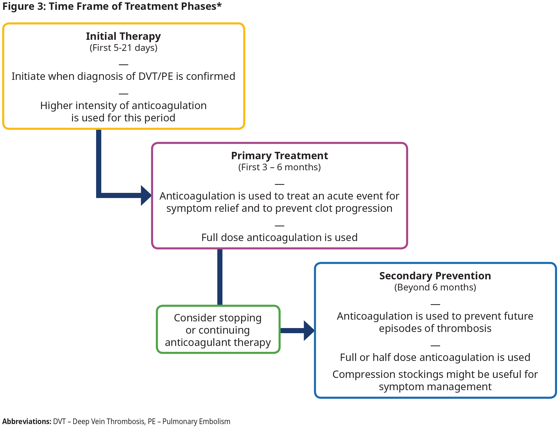
Treatment of DVT/PE is typically considered in different phases that correspond to the likelihood of thrombosis recurrence and intensity (i.e., dosing) of anticoagulation needed. The terminology for these phases varies. The guideline uses terminology outlined in Figure 3: Time Frame of Treatment Phases.
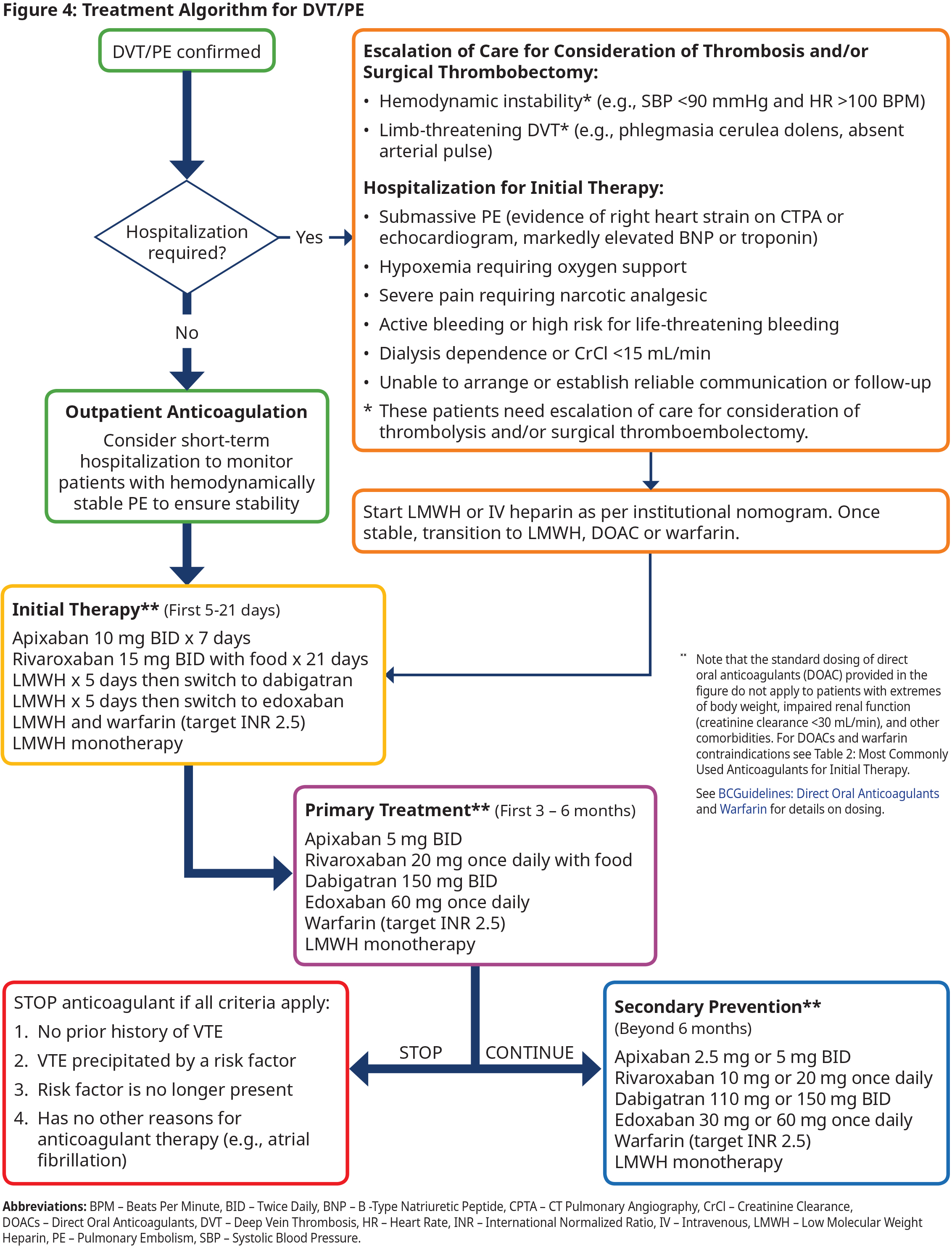
The cornerstone of DVT/PE treatment is anticoagulant therapy. Currently, the most frequently used options include DOACs (mainly apixaban or rivaroxaban), warfarin, LMWH, and intravenous unfractionated (IV) heparin. Each have strengths and limitations, and the most appropriate anticoagulant depends on the clinical scenario, patient factors, and resources available. Refer to BC Guidelines: Direct Oral Anticoagulants and BC Guidelines: Warfarin.
Initial Therapy (first 5-21 days after diagnosis)
- Most patients with DVT and many patients with hemodynamically stable PE can be managed on an outpatient basis. This includes patients with subsegmental or incidental PE11. However, most patients with PE will benefit from short period of observation in the emergency department or hospital admission to allow risk stratification and monitor for hemodynamic stability.
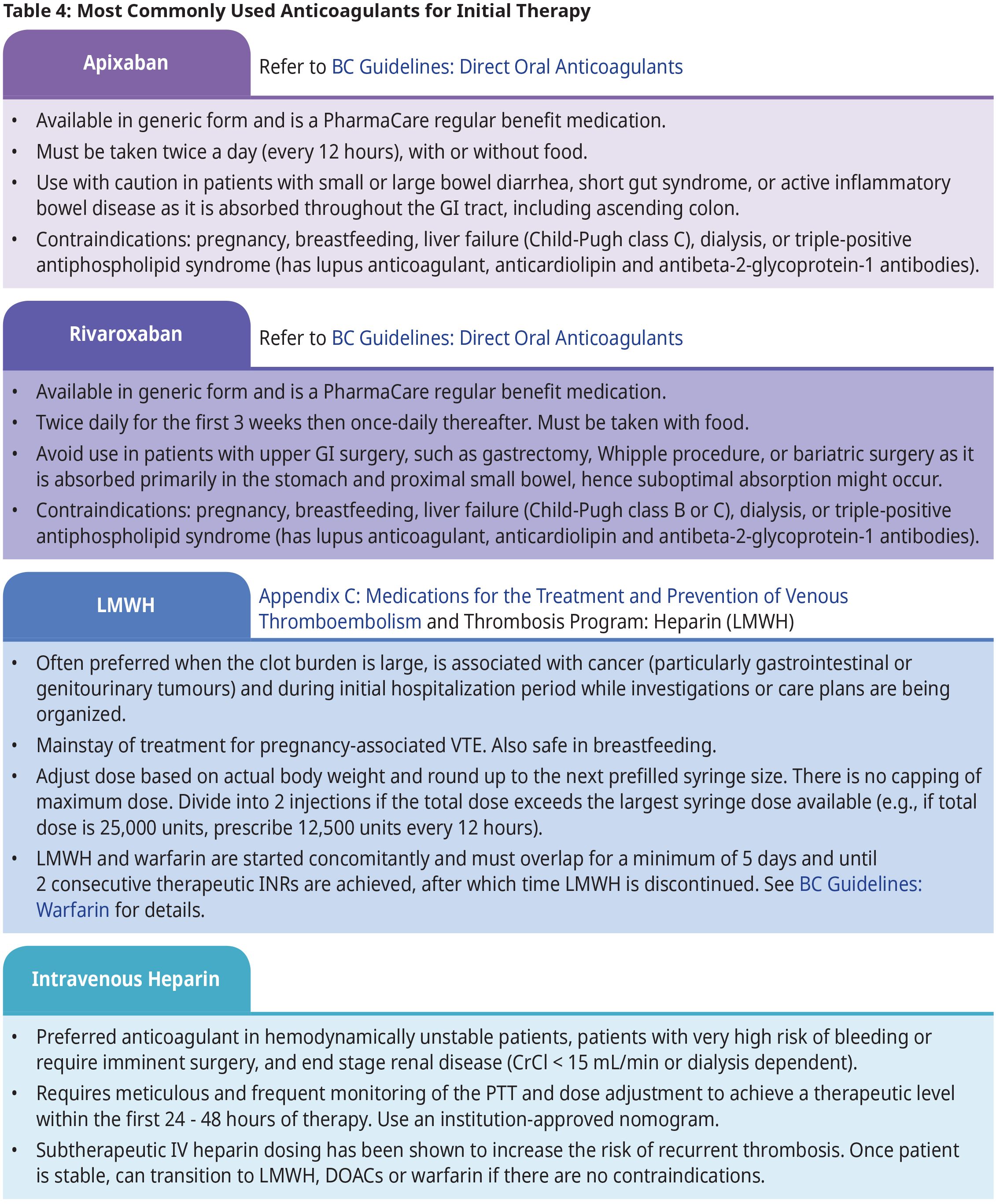
- Initial treatment should be with an immediate acting anticoagulant (onset of action within 2-3 hr), such as apixaban, rivaroxaban, LMWH or IV heparin. See Table 4: Most Commonly Used Anticoagulants for Initial Therapy.
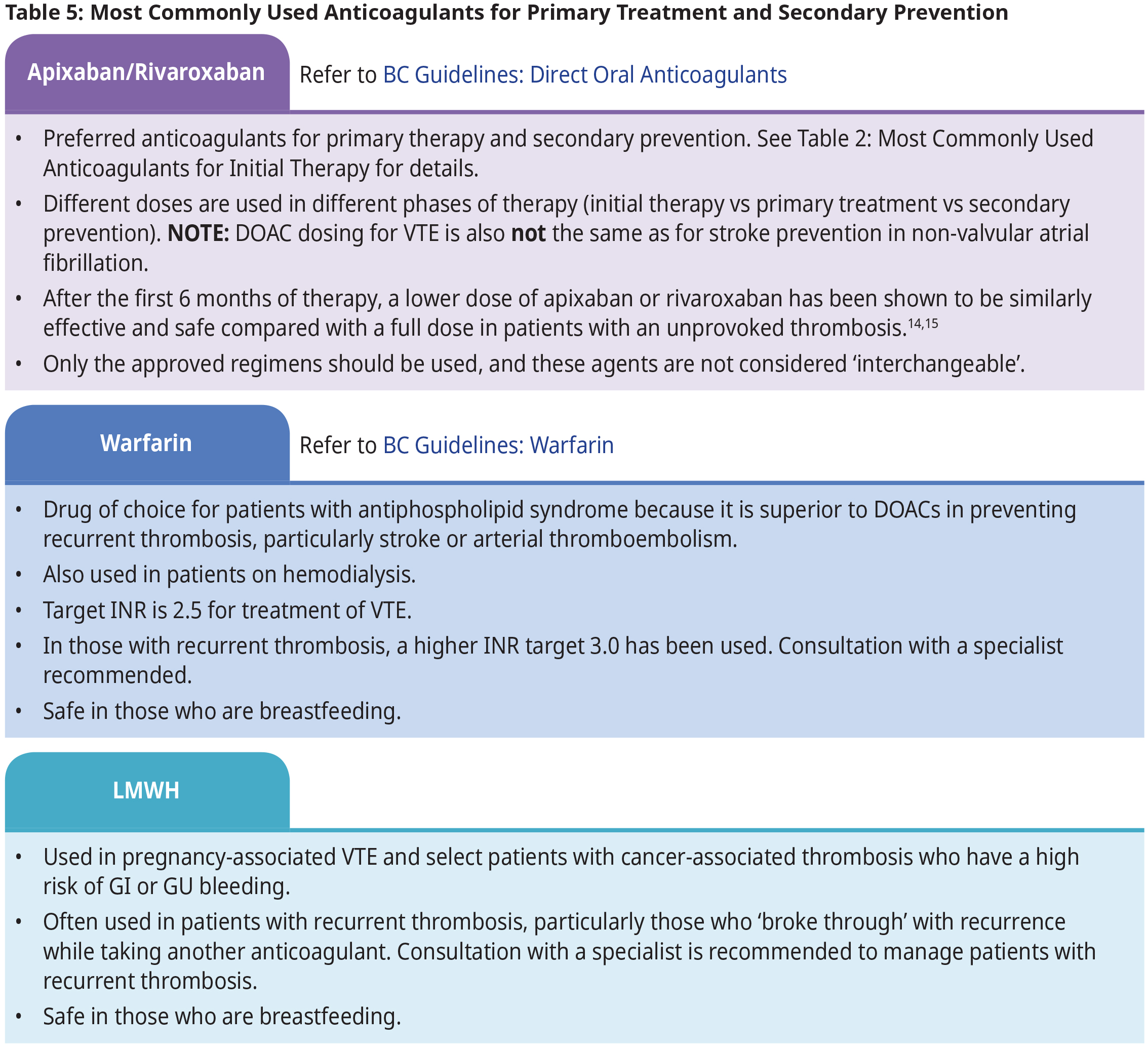
- Apixaban or rivaroxaban are the first-line anticoagulants for most non-pregnant patients because they are effective, safe, and convenient. Indirect evidence suggests patients treated with apixaban experienced a lower rate of recurrent VTE and lower rate of bleeding as compared to rivaroxaban1,2. The choice between apixaban and rivaroxaban depends on practical and pharmacological considerations. See Table 5: Most Commonly Used Anticoagulants for Primary Treatment and Secondary Prevention.
- For patients who cannot be therapeutically anticoagulated due to active bleeding or very high bleeding risks, urgent consultation should be initiated with a hematologist or thrombosis specialist through the RACE line. Management may include placement of a retrievable inferior vena cava filter (i.e., IVC filter) if therapeutic anticoagulation cannot be safely provided in the acute setting.
Primary Treatment (first 3 – 6 months)
- All patients with an acute VTE episode require at least 3 months of therapeutic anticoagulation. See Table 5: Most Commonly Used Anticoagulants for Primary Treatment and Secondary Prevention.
- Some experts recommend extending treatment up to 6 months (or longer) in patients who presented with a large clot burden (e.g., submassive PE, iliofemoral DVT), who remain symptomatic at 3 months, or whose risk factor is still present at 3 months (e.g., cancer).
- Patients are at high risk of recurrent thrombosis during the first 3 months. Elective surgery or procedures that require interruption of anticoagulation should be avoided during this period.
Secondary Prevention (after the first 6 months)
- In general, patients with a first episode of thrombosis that was triggered by a risk factor (e.g., surgery) can stop anticoagulant therapy after 3-6 months, if the risk factor is no longer present after completing primary treatment. The risk of recurrence is typically < 3-5% in these patients.
- Patients without any clear precipitating factor (i.e., unprovoked VTE) are more likely to develop VTE recurrence and may benefit from continuing anticoagulation beyond 6 months. Risk of recurrence in patients with unprovoked VTE is approximately 10% in the first year, 25% in the first 5 years, and up to 40% in the first 10 years after stopping anticoagulant therapy.
- Referral to a hematologist or thrombosis specialist (see BC Thrombosis Network for thrombosis clinics in BC and referral information), especially for those with unprovoked VTE, is recommended to determine if anticoagulation beyond 6 months is indicated, considering the risks of recurrence versus bleeding. While awaiting referral, continuing anticoagulation is recommended.
- Risk of recurrence depends on the precipitating factor(s), the persistence of risk factors (e.g., cancer), physiological predisposition (e.g., age, sex, hereditary thrombophilia), and how strongly each factor promotes clotting (see Table 1: Common Clinical Risk Factors for Venous Thromboembolism).
- Risk of bleeding is multifactorial and increases with the number of risk factors present. Important factors to consider include age >70, active cancer, anemia, concomitant antiplatelet therapy, chronic kidney disease (CKD), chronic liver disease, prior history of bleeding, and thrombocytopenia (platelet count < 50 x 109/L).
- Work-up for occult cancer in patients with unprovoked VTE is not indicated beyond the usual age appropriate malignancy screening.1,3
- Hereditary thrombophilia testing is not indicated for most patients with thrombosis because it does not often influence choice or duration of anticoagulant therapy and is costly.
Supportive Measures
Compression stockings
- Graduated compression stockings are useful for reducing venous congestion symptoms such as edema and muscle tightness or aching. They do not reduce the risk of recurrent thrombosis.
- Over the counter knee-high options (i.e., less than 20 mm Hg pressure) are easy to use and often helpful, particularly for some patients with varicose veins. Prescription grade stockings (i.e., 30-40 mmHg pressure) are often necessary for patients with post thrombotic syndrome (PTS).
- Poorly fitted stockings can cause discomfort and skin breakdown. Tourniquet effect below the knee might cause even more venous stasis and promote thrombosis.
IVC filters
- Filters are sometimes placed in patients with high-risk thrombosis who are unable to receive anticoagulant therapy due to active or high risk of bleeding.
- They should be removed as soon as anticoagulant therapy is safely initiated. Prolonged placement reduces the likelihood of successful retrieval.
- Although evidence is limited, anticoagulation is typically continued if a filter remains in-situ because it is a known cause of recurrent thrombosis.
Health Behaviours
- Foods and supplements cannot be used as an alternative to anticoagulants.
- While taking anticoagulants, patients should avoid herbal supplements due to drug-drug interactions (e.g., St. John’s Wort) and others that can increase the bleeding risk (e.g., ginkgo, garlic, and turmeric)16,17.
- Regular exercise and maintaining a healthy body weight are important behaviours overall and might mitigate the risk of recurrent thrombosis because sedentary lifestyle and obesity contribute to thrombosis formation.
Special populations
Pregnancy
- Thrombosis risk is higher during pregnancy and the first 6-12 weeks postpartum.
- LMWH is the mainstay of treatment for pregnancy associated VTE. Management requires coordination of multiple specialists including hematology/thrombosis, obstetrics, and anesthesia.
- Either LMWH or warfarin can be used in the postpartum period, as neither crosses over into breastmilk.
- DOACs are contraindicated in pregnancy and breastfeeding.
Cancer
- Clinical practice guidelines recommend using LMWH, apixaban, or rivaroxaban for the treatment of cancer associated thrombosis.
- Some DOACs are associated with an increased risk of bleeding in certain malignancies (i.e., intraluminal GI or urothelial tumour site) when compared to LMWH.
- Subspecialist consultation is advised to guide management of cancer-associated thrombosis.
Chronic Kidney Disease
- Patients with CKD are at increased risks of thrombosis and bleeding.
- Patients with stage 4 CKD (i.e., CrCl < 30 mL/min) were excluded from many clinical drug trials so are typically treated with warfarin. Low-dose DOAC is being evaluated in this setting and LMWH (dose adjusted based on anti-Xa level) has also been used.
- Subspecialist consultation (including nephrologist) is advised to guide management.
Antiphospholipid Antibody Syndrome
- APS is an acquired thrombophilia that increases the risk of venous and/or arterial thrombosis events, as well as pregnancy-associated morbidity. Those with triple positivity (has lupus anticoagulant, IgG or IgM anticardiolipin antibodies and IgG or IgM antibeta-2-glycoprotein-1 antibodies) are at highest risk.
- The diagnosis of APS should be made by a specialist due to complex and changing laboratory and clinical diagnostic criteria.
- Treatment of APS-related thrombosis should also be in consultation with a specialist. In general, LMWH and warfarin are the mainstay of therapy in APS. DOACs should be avoided in APS due to weak evidence of efficacy in preventing recurrent (particularly arterial) thrombosis.
Heparin-Induced Thrombocytopenia (HIT)
- HIT is a very rare complication from unfractionated heparin (UFH) or LMWH use that can result in fatal thromboembolism. The onset of thrombocytopenia occurs day 5-14 after first exposure to heparin.
- Consult a specialist immediately if HIT is suspected or confirmed. Heparin should be discontinued immediately and alternate anticoagulation (e.g., fondaparinux or argatroban or DOACs) should be initiated.
Unusual site (splanchnic vein, cerebral vein, upper extremity DVT)
Thrombosis in these sites accounts for approximately 10% of VTE. The optimal therapeutic approach is uncertain due to lack of high-quality evidence. Certain sites of thrombosis may be associated with specific disease states (i.e., splanchnic vein thrombosis in cirrhosis, upper extremity DVT associated with central venous catheters or in thoracic outlet syndrome, cerebral vein thrombosis from OCP use or pregnancy). The management of unusual site thrombosis should be guided by subspecialty expertise.
Follow Up
Most patients will experience improvement in leg and respiratory symptoms within 48-72 hours. Pleuritic chest pain from pulmonary infarction typically lasts a week or longer. By one month, many patients will feel much better and by 3-6 months, most patients will be almost back to baseline. Approximately 1/3 of patients experience residual swelling and/or discomfort in the affected limb (See Thrombosis Canada: Post-thrombotic Syndrome)
There is no risk to exercising or returning to usual activities while on anticoagulation. Exercise is encouraged and patients should be as active as their symptoms allow. The only activities to avoid are contact sports and other activities that put the patient at a higher risk for serious injuries, especially intracranial hemorrhage.
Patients on long-term anticoagulation (i.e., >6 months) should be reviewed annually to determine if continuing anticoagulation remains beneficial. This requires assessing the risks of recurrent thrombosis vs the risk of serious bleeding. See BC Guidelines: Direct Oral Anticoagulants and BC Guidelines: Warfarin for long-term monitoring requirements.
After completion of primary treatment (3-6 months), repeating imaging may be considered on a case-by-case basis per thrombosis specialist recommendation. Note that 1/3 patients will have residual clot on imaging and the presence of residual clot does not determine the duration of anticoagulation.
Resources
Abbreviations
ACLS
APS
BID
BNP
BPM
CHF
CKD
COPD
CPTA
CrCl
CTPA
DOACs
DVT
HIT
HR
INR
IV
IVC Filter
LMWH
PE
SBP
UFH
V/Q
VTE
Advanced Cardiovascular Life Support
Antiphospholipid Antibody Syndrome
Twice Daily
B -Type Natriuretic Peptide
Beats Per Minute
Chronic Heart Failure
Chronic Kidney Disease
Chronic Obstructive Pulmonary Disease
CT Pulmonary Angiography Scan
Creatinine Clearance
Computer Tomography Pulmonary Angiogram
Direct Oral Anticoagulants
Deep Vein Thrombosis
Heparin-Induced Thrombocytopenia
Heart Rate
International Normalized Ratio
Intravenous
Inferior Vena Cava Filter
Low Molecular Weight Heparin
Pulmonary Embolism
Systolic Blood Pressure
Unfractionated Heparin
Ventilation-Perfusion
Venous Thromboembolism
Practitioner Resources
- BC Thrombosis Network: comprises a community of Thrombosis Specialists dedicated to the care of patients with venous thromboembolic disorders. See www.bcthrombosisnetwork.ca/
- RACE Line: Rapid Access to Consultative Expertise Program: raceconnect.ca/. A phone consultation line for physicians, nurse practitioners and medical residents. If the relevant specialty area is available through your local RACE line, please contact them first.
- Pathways: An online resource that allows GPs and nurse practitioners and their office staff to quickly access current and accurate referral information, including wait times and areas of expertise, for specialists and specialty clinics. See: https://pathwaysbc.ca/login
- Health Data Coalition: An online, physician-led data sharing platform that can assist you in assessing your own practice in areas such as chronic disease management or medication prescribing. HDC data can graphically represent patients in your practice with chronic diseases in a clear and simple fashion, allowing for reflection on practice and tracking improvements over time. See: Health Data Coalition – Better Information. Better Care. Better Patient Outcomes. (hdcbc.ca)
Patient, Family and Caregiver Resources
- HealthLinkBC: You may call HealthLinkBC at 8-1-1 toll-free in B.C., or for the deaf and the hard of hearing, call 7-1-1. You will be connected with an English-speaking health-service navigator, who can provide health and health-service information and connect you with a registered dietitian, exercise physiologist, nurse, or pharmacist. See: healthlinkbc.ca/
- Thrombosis Canada: Patient Resources
Diagnostic Codes
I26 - I28 Pulmonary Embolism
I82 Deep Vein Thrombosis
Billing Codes
415.1 Pulmonary Embolism
453 Other Venous Embolism And Thrombosis
451 Deep Vein Thrombosis
452 Portal Vein Thrombosis
Appendices
- Appendix A: Proximal and Distal DVT
- Appendix B: D-dimer Cut-off values
- Appendix C: Medications for the Treatment and Prevention of Venous Thromboembolism
Associated Documents
References
- Jackson CD, Cifu AS, Burroughs-Ray DC. Antithrombotic Therapy for Venous Thromboembolism. JAMA. 2022 Jun 7;327(21):2141.
- Ortel TL, Neumann I, Ageno W, Beyth R, Clark NP, Cuker A, et al. American Society of Hematology 2020 guidelines for management of venous thromboembolism: treatment of deep vein thrombosis and pulmonary embolism. Blood Adv. 2020 Oct 13;4(19):4693–738.
- Heit JA. Epidemiology of venous thromboembolism. Nat Rev Cardiol. 2015 Aug;12(8):464–74.
- Di Nisio M, van Es N, Büller HR. Deep vein thrombosis and pulmonary embolism. The Lancet. 2016 Dec 17;388(10063):3060–73.
- Kearon C. Natural History of Venous Thromboembolism. Circulation. 2003 Jun 17;107(23_suppl_1):I–22.
- Rogers MAM, Levine DA, Blumberg N, Flanders SA, Chopra V, Langa KM. Triggers of Hospitalization for Venous Thromboembolism. Circulation. 2012 May;125(17):2092–9.
- Anderson F, Spencer F. Risk Factors for Venous Thromboembolism. Circulation. 2003 Jul 1;107:I9-16.
- Kearon C, Ageno W, Cannegieter SC, Cosmi B, Geersing GJ, Kyrle PA, et al. Categorization of patients as having provoked or unprovoked venous thromboembolism: guidance from the SSC of ISTH. J Thromb Haemost. 2016 Jul;14(7):1480–3.
- Khan F, Tritschler T, Kahn SR, Rodger MA. Venous thromboembolism. Lancet. 2021 Jul 3;398(10294):64–77.
- Maughan BC, Frueh L, McDonagh MS, Casciere B, Kline JA. Outpatient Treatment of Low-risk Pulmonary Embolism in the Era of Direct Oral Anticoagulants: A Systematic Review. Academic Emergency Medicine. 2021;28(2):226–39.
- Le Gal G, Kovacs MJ, Bertoletti L, Couturaud F, Dennie C, Hirsch AM, et al. Risk for Recurrent Venous Thromboembolism in Patients With Subsegmental Pulmonary Embolism Managed Without Anticoagulation. Ann Intern Med. 2022 Jan 18;175(1):29–35.
- Dawwas GK, Leonard CE, Lewis JD, Cuker A. Risk for Recurrent Venous Thromboembolism and Bleeding With Apixaban Compared With Rivaroxaban: An Analysis of Real-World Data. Ann Intern Med. 2022 Jan 18;175(1):20–8.
- Carrier M, Lazo-Langner A, Shivakumar S, Tagalakis V, Zarychanski R, Solymoss S, et al. Screening for Occult Cancer in Unprovoked Venous Thromboembolism. N Engl J Med. 2015 Aug 20;373(8):697–704.
- Apixaban for Extended Treatment of Venous Thromboembolism | NEJM [Internet]. [cited 2023 Jan 9]. Available from: https://www.nejm.org/doi/full/10.1056/nejmoa1207541
- Weitz JI, Lensing AWA, Prins MH, Bauersachs R, Beyer-Westendorf J, Bounameaux H, et al. Rivaroxaban or Aspirin for Extended Treatment of Venous Thromboembolism. New England Journal of Medicine. 2017 Mar 30;376(13):1211–22.
- Grześk G, Rogowicz D, Wołowiec Ł, Ratajczak A, Gilewski W, Chudzińska M, et al. The Clinical Significance of Drug–Food Interactions of Direct Oral Anticoagulants. Int J Mol Sci. 2021 Aug 8;22(16):8531.
- Clinical Guides [Internet]. Thrombosis Canada - Thrombose Canada. 2013 [cited 2023 Mar 6]. Available from: https://thrombosiscanada.ca/clinicalguides/
Disclaimer:
BC Guidelines are developed for the Medical Services Commission by the Guidelines and Protocols Advisory Committee, a joint committee of Government and the Doctors of BC. BC Guidelines are adopted under the Medicare Protection Act and, where relevant, the Laboratory Services Act.
This guideline is based on best available scientific evidence and clinical expertise as of February 29, 2024. It is not intended as a substitute for the clinical or professional judgment of a health care practitioner.

 TOP
TOP